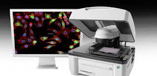
Automatic cell imaging system effectively helps in determining the chromosomal abnormalities in cells
Automatic cell imaging system is the most popular form of noninvasive evaluation for detecting diseases. Since it is noninvasive and can be performed in a laboratory in a fraction of the time it would take in a doctor's office, professionals are continually searching for ways to evaluate tissue samples quickly. Medical professionals have been exploring the idea of using cellular information to assess individual cell function for years. Automatic cell imaging system is playing critical role in the healthcare industry and also across various clinical research applications
Automatic cell imaging system will provide them, such as faster diagnosis, better health monitoring, and less down time due to less patient traffic. By providing a non-invasive alternative to x-rays and mammograms, the technology allows for fewer patient visits. Many of the systems are designed to perform more than one function. For example, the these cell imaging system can be used to inspect mobile phone screens, display damaged parts of the device, diagnose the problem, and fix it automatically.
There is also increasing popularity of the Diagnostic Laboratory software solutions. Many companies manufacture and sell their own diagnostic lab software systems. In order to increase their market share, many manufacturers are introducing products that incorporate market-analysis tools. As diagnostic laboratory software is becoming a popular choice of users across the globe, diagnostic laboratory equipment and accessories are also increasing in popularity. This increase in popularity is due to the need to perform various tests on a regular basis, reliable data transfer capabilities, and fast data retrieval.
Recently, Sysmex Corporation based in Japan has launch the Imaging Flow Cytometer MI-1000 and related software MI FISH Master as a system. This system utilizes imaging FCM technology1 to automate FISH testing2 for determining chromosomal abnormalities in circulating cells.


Comments
Post a Comment